Project Lead(s): Malena Correa
Issue
Pneumonia is the main cause of death due to infection among children under five years of age, worldwide.
It affects disproportionately the poorest, under-served regions, where important barriers in access to proper diagnostic, care and treatment remain as impediments.
Pneumonia diagnosis relies on clinical integration of physical exam, chest X-rays (CXR) and laboratory tests.
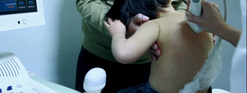
Thoracic ultrasound for lung assessment is becoming a useful and readily available technique for physicians, which has the advantages of being harmless, non-radiating, and time- and cost-saving when compared to CXR and thoracic CT scans.
However, ultrasound still requires costly equipment and trained healthcare personnel to perform and interpret the results, and none of these are available in low-resource settings.
Solution
This project proposed to develop an algorithm that could be used by a neural network – a form of artificial intelligence – into a computer to process and analyze data from a conventional, clinical ultrasound device, to give an easy-to-interpret result to indicate the presence of pulmonary infiltrates (pneumonia).
Data were collected from eight children attending a tertiary hospital in Lima, Peru, who were diagnosed as having pneumonia; and from 30 children attending the same hospital without any respiratory or cardiac complaints (both conditions that may lead to fluid accumulation in the lungs, which could be interpreted as pneumonia infiltrates).
Following a standardized procedure, a physician conducted a complete examination from 12 regions in the thorax, four frontal, four posterior and four lateral, using a clinical ultrasound device.
At least one video was recorded from each region, and these were processed into frames, each frame being further analyzed to generate ‘vector’ (vertical region of a frame). These vectors were classified as positive (lung consolidate) or negative (clean lung, ribs) and analyzed by a computer according to intensity.
A subset of these vectors was used to train the neural network to identify pulmonary infiltrates, to differentiate them from the normal, healthy lung, and to develop an algorithm. At a later stage, another independent subset of vectors, coming from different images, was used to test the computer system and how the algorithm performed.
Outcome
An algorithm was developed capable of identifying the presence of pneumonia with a sensitivity of 91% and a specificity of 100%.
The system can be adapted to smartphones to run in real time while performing the ultrasound examination and, in so doing, allowing an automatized diagnosis immediately and at the point of care.
A small, mobile prototype device – the LOCONIUS (low-cost, non-imaging ultrasound) – was developed, consisting of a box-shaped core with a small computer (or electronic card) inside, and a probe or transducer to produce the ultrasound waves to be analyzed to detect the presence/absence of pneumonia.
The research team has received additional funding as follows: $500,000 from NIH Fogarty International Center; $38,000 US from Pontifical Catholic University of Peru (PUCP) and $76,000 US from CONCYTEC.
The project has been disseminated throughout the scientific community.
The team plans to apply for Phase II Transition To Scale funding to further the development of the final LOCONIUS device.